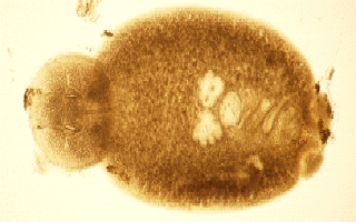
If the surface of the eye is opaque, scrape with a clean scapel blade and place tissues into a drop of saline on a clean glass slide. Cover with a slip cover. Examine under 40 to 400X magnification.
Compare your sample with the following Disease Vectors:
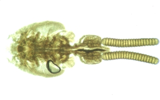
Caligus sp. a parasitic copepod 2 to 3 mm in length
(visable to the naked eye)
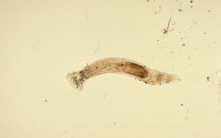
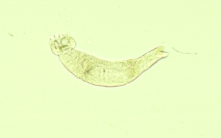
Monogenetic Trematodes parasitic
flatworms. May be small (0.3 to 0.4 mm) or large (2to 3 mm) . Hooks on one
end may be attached to the host fish. If still alive, will flex rapidly or
contract slowly.

| file: /Techref/other/pond/tilapia/eyes.htm, 1KB, , updated: 2018/10/18 01:47, local time: 2025/9/3 19:00,
216.73.216.174,10-3-63-162:LOG IN
|
| ©2025 These pages are served without commercial sponsorship. (No popup ads, etc...).Bandwidth abuse increases hosting cost forcing sponsorship or shutdown. This server aggressively defends against automated copying for any reason including offline viewing, duplication, etc... Please respect this requirement and DO NOT RIP THIS SITE. Questions? <A HREF="http://www.ecomorder.com/techref/other/pond/tilapia/eyes.htm"> Tilapia Topic: Eye Microscopy</A> |
| Did you find what you needed? |
Welcome to ecomorder.com! |
|
The Backwoods Guide to Computer Lingo |
.